National Institute of Information and Communications Technology
Advanced ICT Research Institute
National Institute of Information and Communications Technology
Advanced ICT Research Institute
Uehara T, Yamasaki T, Okamoto T, Koike T, Kan S, Miyauchi S, Kira JI, and Tobimatsu S. Efficiency of a "Small-World" Brain Network Depends on Consciousness Level: A Resting-State fMRI Study. Cerebral Cortex, 2013
article link
Abstract: It has been revealed that spontaneous coherent brain activity during rest, measured by functional magnetic resonance imaging (fMRI), self-organizes a "small-world" network by which the human brain could sustain higher communication efficiency across global brain regions with lower energy consumption. However, the state-dependent dynamics of the network, especially the dependency on the conscious state, remain poorly understood. In this study, we conducted simultaneous electroencephalographic recording with resting-state fMRI to explore whether functional network organization reflects differences in the conscious state between an awake state and stage 1 sleep. We then evaluated whole-brain functional network properties with fine spatial resolution (3781 regions of interest) using graph theoretical analysis. We found that the efficiency of the functional network evaluated by path length decreased not only at the global level, but also in several specific regions depending on the conscious state. Furthermore, almost two-thirds of nodes that showed a significant decrease in nodal efficiency during stage 1 sleep were categorized as the default-mode network. These results suggest that brain functional network organizations are dynamically optimized for a higher level of information integration in the fully conscious awake state, and that the default-mode network plays a pivotal role in information integration for maintaining conscious awareness.
Koike T, Kan S, Misaki M, and Miyauchi S. Connectivity pattern changes in default-mode network with deep non-REM and REM sleep. Neuroscience Research, 69(4): 322-330 (2011)
article link
_Cover.png) Abstract: Recent studies have compared default-mode network (DMN) connectivity in different arousal levels to investigate the relationship between consciousness and DMN. The comparison between the DMN in rapid eye movement (REM) sleep with that in non-REM (NREM) sleep is useful for revealing the relationship between arousal level and DMN, because the arousal level is at its lowest during deep NREM, while during REM sleep it is as high as wakefulness. Functional magnetic resonance imaging (fMRI) and polysomnogram data were acquired from participants in REM, deep NREM, and light NREM sleep, and the DMN was compared using functional connectivity analysis. Our analysis revealed that functional connectivity among the DMN core regions -- the posterior cingulate cortex, rostral anterior cingulate cortex, and inferior parietal lobule -- remained consistent across sleep states. In contrast, connectivity involving the DMN subsystems of REM sleep differs from that of NREM sleep, and the change well accounts for the characteristics of REM sleep. Our results suggest that both the DMN core region and subsystems may not relate to the maintenance of arousal. The DMN core network and subsystems may respectively serve to integrate brain regions and perform function specific to each level of arousal.
Abstract: Recent studies have compared default-mode network (DMN) connectivity in different arousal levels to investigate the relationship between consciousness and DMN. The comparison between the DMN in rapid eye movement (REM) sleep with that in non-REM (NREM) sleep is useful for revealing the relationship between arousal level and DMN, because the arousal level is at its lowest during deep NREM, while during REM sleep it is as high as wakefulness. Functional magnetic resonance imaging (fMRI) and polysomnogram data were acquired from participants in REM, deep NREM, and light NREM sleep, and the DMN was compared using functional connectivity analysis. Our analysis revealed that functional connectivity among the DMN core regions -- the posterior cingulate cortex, rostral anterior cingulate cortex, and inferior parietal lobule -- remained consistent across sleep states. In contrast, connectivity involving the DMN subsystems of REM sleep differs from that of NREM sleep, and the change well accounts for the characteristics of REM sleep. Our results suggest that both the DMN core region and subsystems may not relate to the maintenance of arousal. The DMN core network and subsystems may respectively serve to integrate brain regions and perform function specific to each level of arousal.
Kan S, Koike T, Misaki M, and Miyauchi S. Close relationship between fMRI signals and transient heart rate changes accompanying K-complex: Simultaneous EEG/fMRI study. Japanese Journal of Neurosphysiology, 37(6): 413-422 (2009)
Abstract: Combining functional magnetic resonance imaging (fMRI) and electroencephalography (EEG) allows the investigation of spontaneous activities in the human brain. Recently, by using this technique, increases in fMRI signal accompanying transient EEG activities such as sleep spindles and slow waves were reported. Although these fMRI signal increases appear to arise as a result of the neural activities being reflected in the EEG, when the influence of physiological activities upon fMRI signals are taken into consideration, it is highly controversial that fMRI signal increases accompanying transient EEG activities reflect actual neural activities. In the present study, we conducted simultaneous fMRI and polysomnograph recording of 18 normal adults, to study the effect of transient heart rate changes after a K-complex on fMRI signals. Significant fMRI signal increase was observed in the cerebellum, the ventral thalamus, the dorsal part of the brainstem, the periventricular white matter and the ventricle (quadrigeminal cisterm). On the other hand, significant fMRI signal decrease was observed only in the right insula. Moreover, intensities of fMRI signal increase that was accompanied by a K-complex correlated positively with the magnitude of heart rate changes after a K-complex. Previous studies have reported that K-complex is closely related with sympathetic nervous activity and that the attributes of perfusion regulation in the brain differ during wakefulness and sleep. By taking these findings into consideration, our present results indicate that a close relationship exists between a K-complex and the changes in cardio- and neurovascular regulations that are mediated by the autonomic nervous system during sleep; further, these results indicate that transient heart rate changes after a K-complex can affect the fMRI signal generated in certain brain regions.
Miyauchi S, Misaki M, Kan S, Fukunaga T, and Koike T. Human brain activity time-locked to rapid eye movements during REM sleep. Experimental Brain Research, 192(4): 657-667 (2009)
article link (open access)
Abstract: To identify the neural substrate of rapid eye movements (REMs) during REM sleep in humans, we conducted simultaneous functional magnetic resonance imaging (fMRI) and polysomnographic recording during REM sleep. Event-related fMRI analysis time-locked to the occurrence of REMs revealed that the pontine tegmentum, ventroposterior thalamus, primary visual cortex, putamen and limbic areas (the anterior cingulate, parahippocampal gyrus and amygdala) were activated in association with REMs. A control experiment during which subjects made self-paced saccades in total darkness showed no activation in the visual cortex. The REM-related activation of the primary visual cortex without visual input from the retina provides neural evidence for the existence of human ponto-geniculo-occipital waves (PGO waves) and a link between REMs and dreaming. Furthermore, the time-course analysis of blood oxygenation level-dependent responses indicated that the activation of the pontine tegmentum, ventroposterior thalamus and primary visual cortex started before the occurrence of REMs. On the other hand, the activation of the putamen and limbic areas accompanied REMs. The activation of the parahippocampal gyrus and amygdala simultaneously with REMs suggests that REMs and/or their generating mechanism are not merely an epiphenomenon of PGO waves, but may be linked to the triggering activation of these areas.
Kobashi S, Yahata Y, Kan S, Misaki M, Koike T, Kondo K, Miyauchi S, and Hata Y. Eye Position Estimation During Sleep Using Infrared Video in Functional MRI. Journal of Advanced Computational Intelligence and Intelligent Informatics, 12(1): 32-40 (2008)
(Collaboration with Dr. Kobashi at University of Hyogo)
article link
Abstract: The determination of eye position during sleep attracted much attention in sleep study. Simulaneous measurement consisting of functional magnetic resonance imaging and infrared video was developed to study the relationship between eye movement and brain function during sleep. Conventional measurement of the eye position using infrared video images is not applicable to experiments during sleep because of the lack of tracking targets such as the pupil. This paper proposes a novel method for determining the eye position during sleep through infrared video images. This method determines the eye position by comparing intensity profile extracted from an image to intensity profiles generated by the artificial neural network (ANN). Experiments showed that the proposed method detected the eye position of the left eye with an error of 3.56 +/- 3.61 (RMSE +/- SD) pixels and that of the right eye with an error of 4.69 +/- 4.73 pixels.
Misaki M, Miyauchi S. Application of artificial neural network to fMRI regression analysis. NeuroImage, 29(2): 396-408 (2006)
article link
Abstract: We used an artificial neural network (ANN) to detect correlations between event sequences and fMRI (functional magnetic resonance imaging) signals. The layered feed-forward neural network, given a series of events as inputs and the fMRI signal as a supervised signal, performed a non-linear regression analysis. This type of ANN is capable of approximating any continuous function, and thus this analysis method can detect any fMRI signals that correlated with corresponding events. Because of the flexible nature of ANNs, fitting to autocorrelation noise is a problem in fMRI analyses. We avoided this problem by using cross-validation and an early stopping procedure. The results showed that the ANN could detect various responses with different time courses. The simulation analysis also indicated an additional advantage of ANN over non-parametric methods in detecting parametrically modulated responses, i.e., it can detect various types of parametric modulations without a priori assumptions. The ANN regression analysis is therefore beneficial for exploratory fMRI analyses in detecting continuous changes in responses modulated by changes in input values.
Miyauchi S, Kan S, Koike T, and Misaki M. Simultaneous EEG/fMRI recording and resting-state fMRI for investigation of brain state. 16th Annual Meeting of the Japanese Society of Cognitive Neuroscience, Kitakyushu. 22 Oct. 2011
Kan S, Koike T, and Miyauchi S. The activation of the pons accompanying rapid eye movements during REM sleep: a simultaneous EEG/fMRI study. 13th Congress of Japan Human Brain mapping Society, Kyoto. 1 Sep. 2011
Abstract: Using simultaneous EEG/fMRI recording, several previous studies have reported brain activities in cortical and subcortical areas accompanying rapid eye movements during REM sleep. However, activations of the brainstem that is closely related to several phenomena during REM sleep are still controversial. Possible cause of such inconsistency is low signal-noise ration of 1.5T MRI that previous studies used. In this study, we investigated brainstem activity accompanying REMs using 3T MRI and high-resolution fMRI scan. Using simultaneous EEg/fMRI recording, we measured spontaneous brain activities from 8 normal adult subjects. We visually identified REM periods and onsets of REMs, and then we conducted event-related analysis with SPM8. In this analysis, we considered REMs as events. Six of eight subjects reached REM sleep. Data from two of them were eliminated further analysis due to artifacts in EPI images. We observed REMs-related activations of primaly visual cortex and the ponse in all four subjects. Location of REMs-related pontine activation corresponded to the paramedian pontine reticular foramtion (PPRF). In contrast, REM-related deactivation was not observed. In this study, we found activation of the PPRF accompnaying REMs during REM sleep. The PPRF is orgin of ponto-geniculo-occipital (PGO) waves that are closely related to generation of REMs. Moreover, the PPRF is involved with eye movements during wakefulness such as optokinetic nystagmus and nicotine-induced nystagmus. Considering these findings, our results suggest that REMs-related activation of the PPRF reflects PGO waves and/or generation of eye movements.
Kan S, Koike T, Misaki M, and Miyauchi S. Resting-state fMRI: investigations of spontaneous brain activities. 26th Annual Meeting of Japan Biomagnetism and Bioelectromagnetics Society, Fukuoka. 4 Jun. 2011
Abstract: In recent years, we can investigate spontaneously organized brain networks with resting-state fMRI and functional connectivity analysis. Using these technique, several studies have reported that these network are related to personal cognitive abilities and several neuropsychiatric disorders. Although there are some issues related to neural basis and functional role of the network, resting-state fMRI and functional connectivity analysis are helpful to investigate brain activities during a variety of unconscious states such as sleep, anesthesia and coma. Recently, we investigated brain networks during light-NREM, deep-NREM and REM sleep, and we observed changes of some connections between the default mode network and other regions. In this presentation, we will introduce our results in detail and discuss pitfalls of resting-state fMRI.
Kan S, Koike T, and Miyauchi S. Activation of the pontine reticular formation accompanying rapid eye movements during REM sleep: A high-resolution fMRI study. 40th Annual Meeting of Society for Neuroscience, San Diego CA, USA. 14 Nov. 2010
Abstract: Numerous animal studies have demonstrated that the brain stem plays a key role in the generation of ponto-geniculo-occipital (PGO) waves and rapid eye movements (REMs) during REM sleep, but the related neural substrate in the generation of these phenomena so far remains unclear in humans. In recent years, we and other groups have reported that several brain areas, such as the thalamus and the visual cortex, are activated accompanying REMs by using fMRI. Among these studies, activation areas in the visual cortex and the thalamus mostly overlap; however activation regions in the brain stem are inconsistent. We investigated brain stem activities accompanying REMs by a high-resolution fMRI at 3T to precisely localize activation regions. Five healthy adults participated in the study. We recorded fMRI and EEG data simultaneously while they slept in an MR scanner. After subtracting scanning and pulse artifacts from the EEG, we visually identified the periods of REM sleep and onsets of REMs from the EEG, EOG and video recordings of participants' eyelid movements. Then we conducted event-related analysis with SPM8. As reported in previous studies, we observed REMs-related activation of the visual cortex and the thalamus. Note that we found additional activation specially localized in the medial part of the pontine tegmentum; likely the pontine reticular formation (PRF). It is well known that the PRF, as well as the pedunculopontine nucleus, is closely related in the generation of PGO waves; however, we assume that the activation of the PRF is associated with the generation of REMs, based on the following findings: (1) activation of the PRF was observed during horizontal optokinetic nystagmus (OKN) in a previous study; (2) a lesion in this area impairs REMs as well as waking saccades. Although our result, consequently, suggests that REMs and waking saccades share a neural substrate underlying oculomotor function, this does not mean REMs are completely equivalent to waking saccades. In our additional control experiment we were not able to observe pronounced pontine activation elicited by horizontal OKN or waking saccades, even when we adopted block design, which has higher detection power than event-related design. In addition, a previous study on cats reported that the slope of main sequence amplitude-velocity relationship of REMs is higher than that of waking saccades, implying that the oculomotor system is in a different state of excitation during REM sleep and wakefulness. Considering these findings, our result suggests that the PRF has a different functional significance in REMs during REM sleep and saccades during wakefulness.
Miyauchi S, Kan S, Koike T, and Misaki M. fMRI activation time-locked to rapid eye movements during REM sleep. 29th International Congress of Clinical Neurophysiology, Kobe. 1 Nov. 2010
Abstract: Frequent eye movement and high incidence of vivid dreams are characteristic of rapid eye movement (REM) sleep. Several studies have reported REM-related neural activities in cortical and subcortical areas, which are inactive during waking saccades in total darkness. However, the exact locations of these areas are still unclear. To identify the neural substrate of rapid eye movements during REM sleep in humans, we conducted simultaneous functional magnetic resonance imaging (fMRI) and polysomnographic recording during REM sleep. Event-related fMRI analysis time-locked to the occurrence of REMs revealed that the activation of the pontine tegmentum, ventroposterior thalamus, primary visual cortex (V1), putamen, and limbic areas (the anterior cingulate, parahippocampal gyrus, and amygdala) occur in association with REMs. The results of a control experiment during which subjects made self-paced saccades in total darkness showed no activation in the visual cortex. The time-course analysis of blood oxygenation leveldependent (BOLD) responses indicated that the activation of the pontine tegmentum, ventroposterior thalamus, and primary visual cortex started before the occurrence of REMs (from 3 to 5 s). In contrast, the activations of the putamen and limbic areas accompanied REMs. The results can be summarized as follows: (1) the REM-related activation of the pontine tegmentum, thalamus, and primary visual cortex without visual input from the retina provides neural evidence for the existence of ponto-geniculo-occipital waves (PGO-waves) in humans; (2) the V1 activation prior to REM is consistent with the scanning hypothesis of dreaming; i.e., dream imagery occurs first, and then the eyes scan it; and (3) the activation of the parahippocampal gyrus and amygdala accompanying REMs implies that REMs essentially function as triggers that activate these memory-related areas rather than mere epiphenomenon of PGO-waves.
Kan S, Koike T, Uehara T, Tobimatsu S and Miyauchi S. The reticular activating system is associated with spontaneous EEG/fMRI study. 29th International Congress of Clinical Neurophysiology, Kobe. 30 Oct. 2010
Abstract: Objective: In recent years, brain activation correlated with spontaneous fluctuations of alpha rhythm during relaxed wakefulness has been intensively investigated using the simultaneous EEG/fMRI recording technique. Whereas a negative correlation between alpha rhythm and BOLD signal in the occipital cortices was consistently observed, positive and negative correlations in other regions are controversial among previous studies. Furthermore, studies that investigated activation of subcortical regions related to alpha rhythm are scarce. In this study, we investigated activation of subcortical structures, related to spontaneous alpha rhythm during resting state, besides cortical regions. Methods: We simultaneously measured EEG and fMRI data from 26 participants (20-35 year-old). During 10-minutes recording sessions, they lay supine in a 3T-MRI scanner and were resting with eye closed. After removing the MR gradient artifacts and the ballistocardiogram artifacts in the EEG data, we calculated relative alpha power (ratio between 8-12 Hz and 1-12 Hz band power) corresponding to each EPI image from 7.5 sec windows of EEG data, which was used as a regressor of interest in individual analysis. In addition, we used realign parameter as a nuisance regressor. Individual and group analysis was done using SPM2. Results: We found a negative correlation in the occipital cortex, which is consistent with previous studies. We also found a negative correlation in the superior temporal gyrus and the sensorimotor cortex. In contrast to the negative correlation, a positive correlation was mainly observed in subcortical regions, such as the thalamus, lateral part of the midbrain tegmentum and medial part of the pontine tegmentum. Conclusions: We found specific subcortical structures related to spontaneous fluctuations of alpha rhythm. These structures seem to correspond to the reticular activating system. Our finding, thus, suggests that reticular activating system is involved in fluctuations of spontaneous alpha rhythm during resting wakefulness.
Kan S, Koike T, Uehara T, Tobimatsu S and Miyauchi S. Influence of convolution of the hemodynamic response function on identification of alpha-fMRI correlations. 33rd Annual Meeting of the Japanese Neuroscience Society for Neuroscience, Kobe. 2 Sep. 2010
Abstract: Objective: Recently, brain activation correlated with spontaneous fluctuations of alpha rhythm during wakefulness has been intensively investigated. Almost these studies used canonical hemodynamic response function (HRF); however, this is not justified yet. To investigate influence of convoluting HRF, we compared results of the following two models in this study: the first model used alpha time series, and the second model used that convolved with HRF.Methods: We simultaneously measured EEG and fMRI data from 26 participants (age: 20-35 year-old). During 10-minutes recording sessions, they lay supine in a 3T-MRI scanner and were resting with eye closed. We calculated relative alpha power (ratio between 8-12 Hz and 1-12 Hz band power) corresponding to each EPI image from 7.5-sec window of EEG data, which was used as a regressor of interest. For the second model, we used another regressor derived from the alpha time series convolved with HRF. In addition, we used realign parameter as a nuisance regressor in both models. Individual and group analysis was done using SPM2.Results: For the first model, we found positive correlation in the thalamus, the midbrain-pontine tegmentum and the cerebellum. We also found negative correlation in the occipital cortex, the sensorimotor cortex and the superior temporal gyrus. In contrast to the first model, we did not observe positive correlation, and we observed negative correlation almost all cortical regions, except for prefrontal areas, for the second model.Conclusions: We demonstrated that convolution of HRF largely affects detection of alpha-fMRI correlations. In contrast to the first model, results of the second model, especially positive correlation, is artifactual, because positive correlation was observed only in the lateral ventricle. Our results clearly show that we must consider validity of convoluting HRF in the future studies exploring neural correlates of spontaneous fluctuations of alpha rhythm.
Kan S, Koike T, Misaki M, and Miyauchi S. Heart rate fluctuation affects fMRI signals across the cerebral cortices during REM and light non-REM sleep, but not during deep non-REM sleep. 39th Annual Meeting of Society for Neuroscience, Chicago IL, USA. 19 Oct. 2009
Abstract: Recently, the effects of cardiac activity, especially heart rate, on fMRI signals have been reported. We previously reported a positive correlation between heart rate and fMRI signals across many cortical areas, including the medial prefrontal and occipital cortices, during light non-REM sleep (sleep stage 2). During light non-REM sleep, a biphasic change in the heart rate occurs accompanied by a K-complex which is a characteristic waveform in sleep EEG. Thus, we assumed that the positive correlation between the 2 parameters may be light non-REM sleep-specific. To test this assumption, we investigated the relationship between the 2 parameters by performing correlation (simple regression) analysis using SPM2 during different sleep stages: REM and light and deep non-REM sleep (sleep stage 3/4). We simultaneously obtained fMRI and polysomnography recordings in 15 participants (age, 22-31 years) while they slept during 2 consecutive nights. Of the 15 participants, 12 entered REM sleep, and 11 and 12 showed stable light and deep non-REM sleep, respectively. In REM sleep, as well as light non-REM sleep, significant positive correlation between the 2 parameters was observed across many cortical areas, including the posterior cingulate and posterior insular cortices. The positive correlation was also observed in both the posterior thalamus and cerebellum. In contrast, in deep non-REM sleep, significant positive correlation was observed only in the occipital cortex and thalamus. Moreover, the positive correlation in the thalamus observed during deep non-REM sleep was significantly higher than that observed during light non-REM and REM sleep. Negative correlation was not observed during any of the sleep stages. Heart rate fluctuation is relatively greater in light non-REM and REM sleep than that in deep non-REM sleep. In addition, the coefficient of variation of R-R intervals (CVRR) at each sleep stage also showed the same tendency in our study. Thus, the positive correlation between the 2 parameters across the cerebral cortices may be due to the greater heart rate fluctuation. On the other hand, the positive correlation in the thalamus may be related to the observed correlation between EEG delta power and regional cerebral blood flow in the thalamus, because the thalamus is associated with the regulation of cardiovascular activity and the generation of EEG delta rhythm. In conclusion, the relationship between heart rate and fMRI signals differs across each sleep stage. Hence, the effect of heart rate fluctuation during sleep should be considered while conducting an fMRI study.
Koike T, KanS, Uehara T, Tobimatsu S, and Miyauchi S. Alterations in brain network corresponding to the fluctuations in arousal level within a short time period. 39th Annual Meeting of Society for Neuroscience, Chicago IL, USA. 21 Oct. 2009
Abstract: [PURPOSE] Several studies have examined the characteristics and spatial patterns of the default mode network (DMN) during resting wakefulness. Arousal levels do not remain constant even for short periods of time. Even when participants claim to have a clear sensorium while being scanned, the electorencephalogram (EEG) pattern indicates that high- and low-arousal microstates alternate with each other. In this study, we attempted to elucidate whether the activity of the DMN brain network fluctuates with changes in arousal level during a relatively short time period. [METHOD] We acquired simultaneous functional magnetic resonance imaging (fMRI) and EEG data from 12 study participants who were required to stay awake and keep their eyes closed while being scanned within the MRI scanner. The arousal level_defined as the ratio between EEG power, rAT = (EEG power in alpha-band (8-12 Hz) / the EEG power in the theta-band (4-8 Hz)) was observed to have significantly fluctuated during the session (10 min). Higher rAT values indicate higher arousal levels. We defined microstates with a rAT>1 as ghigh-arousal microstates,h and microstates with a rAT<=1 as glow-arousal microstates.h Subsequently, using preprocessed fMRI data, we evaluated functional connectivity during high- and low-arousal microstates. Functional connectivity was calculated across 22 regions of interest (ROIs) including subcortical regions, visual cortices, and fundamental elements of DMN using Pearsonfs correlation coefficient. Thereafter, we tested whether the strength of connectivity varied between high- and low-arousal microstates by using Wilcoxonfs matched-pair signed-rank test. [RESULTS and DISCUSSION] First, the positive connectivity between the fundamental elements of the DMN was observed; it was observed that the strength of connectivity significantly increased during the high-arousal microstate as compared to the low-arousal (p < 0.001). To our knowledge, this is the first study showing that DMN characteristics change with short-time fluctuations in the arousal level. In addition, we found that the strength of the anticorrelation between the thalamus and the visual cortex increased during the high-arousal compared to the low-arousal microstates (p = 0.002); positive connectivity within the visual cortex was enhanced during the high-arousal microstates (p = 0.02). These modulations associated with the thalamo-visual network appears to be consistent with the evidence that the EEG alpha-wave originates from the thalamo-visuo circuit. The present study indicates that the functioning of brain networks varies with fluctuations in the arousal levels in resting wakefulness.
Koike T, Kan S, Misaki M, and Miyauchi S. Rapid eye movements and waking saccades share the same brain network with different characteristics. 32nd Annual Meeting of the Japanese Neuroscience Society for Neuroscience, Nagoya. 17 Sep. 2009
Abstract: Using functional magnetic resonance imaging (fMRI), we explored functionally connected brain networks associated with waking saccades (visually guided saccade, VG; self-paced saccade with eyes open/closed; EO/EC) and rapid eye movements (REMs) during REM sleep.Functional connectivity between V1 and saccade-related areas were significantly positive in VG and EO, and almost no connectivity was observed in EC cases. The positive connectivity between V1 and saccade-related areas represents the mechanism for controlling eye movements on the basis of visual information.In the case of REMs, robust but negative connectivity was observed between V1 and saccade-related areas. The results suggest that the same network for the control of eye movements is associated with both waking saccades with visual information processing and REMs; however, the functional role of the network during REM sleep may be different from that during performing waking saccade tasks.
KanS, Koike T, Uehara T, Tobimatsu S, and Miyauchi S. Sleep-specific relationship between heart rate fluctuation and functional magnetic resonance imaging signal across cortical areas. 32th Annual Meeting of the Japanese Neuroscience Society for Neuroscience, Nagoya. 18 Sep. 2009
Abstract: We previously reported a positive correlation between heart rate and fMRI signal across many cortical areas including the medial prefrontal and the occipital cortex during light non-REM sleep. To examine whether this correlation is sleep-specific, we performed investigations at relatively high- and low-arousal wakefulness. In high-arousal wakefulness, in contrast to light non-REM sleep, only negative correlation was observed in the periventricular area. The negative correlation was preserved in low-arousal wakefulness. This result clearly shows that the positive correlation across the cortex is sleep-specific. In light non-REM sleep, K-complex occurs spontaneously or provocatively accompanied by a biphasic change in the heart rate. Thus, such a change in heart rate accompanied by the K-complex may lead, at least partially, to the sleep-specific positive correlation between heart rate and the fMRI signal across the cortex.
Koike T, Kan S, Uehara T, Tobimatsu S, and Miyauchi S. The fluctuation of brain network corresponding to the fluctuation of arousal level within a relatively short time. The 11th Congress of Japan Human Brain Mapping Society, Nigata. 29 May. 2009
Kan S, Koike T, Uehara T, Tobimatsu S, and Miyauchi S. The influence of heart rate fluctuation on fMRI signal is localized to periventricular areas. The 11th Congress of Japan Human Brain Mapping Society, Nigata. 29 May. 2009
Koike T, Kan S, Misaki M, and Miyauchi S. Exploring changes in the brain network related to human sleep states using graph theory. 38th Annual Meeting of Society for Neuroscience, Washington DC, USA. 17 Nov. 2008
Abstract: The focus of our research is on changes in the functional organization of the human brain during the course of REM/NREM sleep. In particular, we examined what brain regions are indispensable for sleep-related network, and how the brain network switches in association with REM/NREM transition. Given several lines of evidence showing the possibility that REM/NREM sleep have different role in information processing, e.g. memory consolidation, there would be at least two different brain networks corresponding to REM/NREM sleep. In order to evaluate switching of brain network, we employed graph theory, which is usually considered as the most appropriate framework to analyze complex networks such as human brain network. We recorded fMRI on thirteen normal subjects while they slept in an MR scanner without any goal-directed tasks. Sleep stages were scored from EEG, EMG, EOG, and video data which were simultaneously recorded with fMRI. First, we modeled the sleeping brain as the sleeping brain as a non-directed weighted graph with continuous arch weights given by the hyperbolic correlation measure between spontaneous BOLD signals at any two different regions of interests. Next, we calculated the vulnerability of the network to identify what brain regions are the most indispensable to the functionally connected brain networks. During REM sleep states, following brain regions were detected as vulnerable regions: the thalamus, putamen, amygdala, and insula cortex. During NREM sleep (SWS; slow-wave-sleep), the most central areas are: the thalamus, putamen, amygdale, and caudal part of hippocampus. Finally, in order to evaluate which brain regions communicate with the vulnerable regions, we evaluated the network efficiency. As a result, it is revealed that hippocampus interacted with association area (BA46, 19, 9, and precuneus) during REM sleep, while hippocampus dominantly communicates with subcortical areas (the thalamus, putamen, amygdale, insula, and caudate) during NREM sleep. The thalamus significantly exchanged information between primary cortices (V1, S1, A1, and M1) only during REM sleep. Other vulnerable regions, i.e. the putamen, amygdala, and insula, equally interacted with cortical areas during both REM and NREM. The result of our experiment is likely the first evidence to clearly show the switching of the functionally connected brain network accompanying REM/NREM transition. The observed switching of information flow between thalamus/hippocampus and other brain regions is consistent with "two stage hypothesis of memory consolidation" proposed by Stickgold et al. (2000).
Kan S, Koike T, Misaki M, and Miyauchi S. Simultaneous EEG/fMRI recording for studying human sleep: Relationship between K-complex and cerebral perfusion autoregulation. 38th Annual Meeting of the Japanese Society of Clinical Neurophysiology, Kobe, Japan. 13 Nov. 2008
Abstract: Although BOLD-fMRI is based on neurovascular coupling, it is unclear whether neurovascular coupling is preserved during sleep. The K-complex, a major graphoelement in sleep EEGs, is followed by changes in heart rate and blood pressure. These cardiovascular changes that accompanied the K-complex may affect the fMRI signal. Thus, we conducted simultaneous fMRI and polysomnograph studies on sleeping participants and attempted to distinguish the component reflecting the heart-rate fluctuation from the fMRI signal change that accompanied the K-complex. The fMRI signal significantly increased in the periventricular white matter, cerebellum, thalamus/hypothalamus, and pons/midbrain, and the signal significantly decreased in the insula and the auditory and visual cortices. Furthermore, the amplitude of the signal change in the periventricular white matter was proportional to the degree of heart-rate fluctuation after the K-complex. These results indicate that the fMRI signal change accompanying the K-complex includes neural as well physiological/vasculative components. Since the arterioles in this region are involved in cerebral perfusion autoregulation and the CO2 reactivity in some brain regions changes during sleep, the fMRI signal change accompanying the K-complex suggests that the K-complex is associated with the control of perfusion autoregulation during sleep.
Koike T, Kan S, Misaki M, and Miyauchi S. Spontaneous BOLD fluctuation analysis based on hierarchical clustering technique identifies REM/NREM neural network. 18th Annual Meeting of Japanese Neural Network Society, Tsukuba, Japan. 24 Sep. 2008
Abstract: Using spontaneous BOLD fluctuation analysis based on hierarchical clustering technique, we tried to identify neural networks corresponding to REM/NREM sleep. We found 2 sub-networks during REM and 3 networks during NREM. The results may suggest different functional roles of REM/NREM.
Koike T, Kan S, Misaki M, and Miyauchi S. Dynamic switching of thalamocortical network with transition of human states between NREM and REM sleep. 13th Annual Meeting of the Organization for Human Brain Mapping, Melbourne, Australia. 19 June 2008
Abstract: Increased attention has been directed at investigating spontaneous BOLD activities, and these studies have made clear that several brain regions may form state-dependent network without any goal directed tasks, and the spontaneous network is thought to correlate with some brain functions. Several recent studies suggest that human sleep is not only a resting state but a state processing important function, such as memory consolidation, and suggest the possibility that the REM and NREM sleep have different function. Therefore, it is reasonable to suppose that state-dependent network switches its characteristics accompanying sleep stage transition between REM and NREM. In this study, using spontaneous BOLD activity analysis, we tried to find out dynamic switching of state-dependent brain network with transition of sleep state.
Koike T, Kan S, Misaki M, and Miyauchi S. Dynamic switching of thalamocortical network in association with transition between REM and NREM sleep. SEIRIKEN KENKYU-KAI: fMRI - a tool for neuroscience research, Okazaki, Japan. 22 Nov. 2007
Abstract: In recent years, increased attention has been directed at investigating spontaneous brain activities; examination of spontaneous BOLD activity has made clear that several brain regions form a functionally- connected "resting-state network" during wakefulness without goal-directed tasks. Another attention has been paid to brain activities during sleep. Although there seems to be also no explicit goal-directed tasks during human REM/NREM sleep state, several recent studies suggest that spontaneous brain activities during sleep are associated with important brain functions such as memory consolidation. Therefore, considering functionally-connected brain network associated with human REM/NREM sleep may be helpful to investigate sleep-stage-related brain functions. To investigate characteristics of spontaneous networks linked to human sleep states, we used fMRI with simultaneous polysomnographic recording, and examined functional connectivity between thalamus and other cortical areas, using a technique with Spearman's rank-correlation coefficient between the BOLD signals. We found that a set of regions, including DLPFC, anterior cingulate and hippocampus, showed significantly increased connectivity to thalamus during NREM sleep period (sleep stage 3/4). In contrast, during REM sleep which is usually linked to vivid dreaming, we found a thalamocortical network including visual cortices, hippocampus and primary motor area. In addition, only during REM sleep, we found a significant functional connectivity between the thalamus and the posterior cingulate cortex, precuneus, and inferior temporal gyrus; these regions are known as a part of "default network". Our findings show that thalamocortical network switches in association with transition of sleep states between NREM and REM, and suggest that identifying functionally-connected brain network using correlation coefficient between BOLD signals is a useful tool for exploring brain functions of NREM and REM sleep.
Misaki M, Abe T, Kan S, and Miyauchi S. Relationship between the EEG rhythm and BOLD signals. 29th Annual Meeting of the Japan Neuroscience Society, Kyoto, Japan. 20 July 2006
Abstract: Recording fMRI and an EEG simultaneously is effective for studying spontaneous brain activities. We used this method to examine the relationship between an EEG rhythm and a BOLD signal. Some studies have hypothesized that the hemodynamic response for a change in power of certain EEG frequency bands, such as alpha waves, is canonical in shape. However, the appropriate response shape for a change in the rhythmic EEG has not yet been determined. We measured the EEG and fMRI simultaneously while subjects were in a resting or sleeping state. We applied nonlinear regression analysis using an artificial neural network to detect correlations between the changes in rhythmic EEG waves and fMRI signals without a priori hypothesis of the response shape.
Miyauchi S, Misaki M, Kan S, and Abe T. Brain activation and its time course accompanying raped eye movements during REM sleep revealed by simultanoeus polysomnographic recording with fMRI. 12th Annual meeting of the Organization for Human Brain Mapping, Florence, Italy. 14 June 2006
Abstract: Despite numerous studies on REM sleep, two issues remain unsettled in this field. One is the lack of clear evidence for PGO-waves in humans; the other is whether there is a relationship between rapid eye movement and dreaming, the so called "scanning hypothesis" according to which REM is directly correlated to the movements of the gaze in a dream scene. Though some EEG/MEG studies found neural activities in the occipital area and the thalamus which accompanied REM, the precise location of these brain areas is still unclear. To address these issues, ten subjects slept in an MR scanner for a few hours in the early morning. We simultaneously obtained an fMRI signal accompanied by polysomnography and video recording of eyelid movements. After removing artifacts caused by the MR scanner from the polysomnograph, we visually identified the period of REM sleep and eye movement onsets using both the polysomnograph and the video recording. Six of the subjects showed stable REM sleep which lasted for 20-40 min. Event-related fMRI analysis was done using SPM2. The expected BOLD response for each REM onset was modeled by a canonical HRF. In addition, other models in which the event onset time was shifted were applied to detect REM-related activities with different latencies. All six of these subjects showed a similar fMRI activation pattern accompanying REM: the pontine tegmentum, thalamus, primary visual cortex, extrastriate cortex, cuneus, cingulated cortex, basal ganglias and cerebellum were significantly activated. The activation of the first three areas probably reflects neural activities caused by PGO-waves. In addition, whereas the activation of the first three areas tended to precede REM onset, the activation of the other areas was delayed by a few seconds. The precedent activation to REM in V1 suggests that dream imagery occurs first, and then eyes move to scan the image as is consistent with the scanning hypothesis.
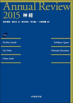 Our review paper entitled "Researches for sleep and consciousness with simultaneous EEG/fMRI" has been published in Annual Reviewe 2015 "Nerve".
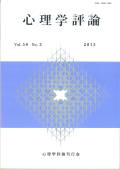 Satoru Miyauchi published a review paper entitled "Measurement of the brain" in Japanese Psychological Review. Vol. 56 No. 3, 2013.
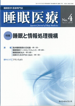 Our review article entitled "fMRI studies for spontaneous and evoked brain activities during sleep" has been published in Japanese Journal of Sleep Medicine. Vol.4 No.4, 2010.
 Our review article entitled "functional connectivity of spontaneous brain activity" has been published in Clinical Neuroscience. Vol.28 No.10, 2010.
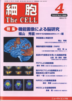 Our review article entitled "functional magnetic resonance imaging (fMRI) for brain science" has been published in The CELL. Vol.41 No.4, 2009.
 Our review article entitled "functional magnetic resonance imaging for sleep studies" has been published in Sleep Research (ed. The Japanese Society of Sleep Research).
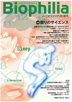 Our review article entitled "Seeing brain activities during sleep: Development of simultaneous functional magnetic resonance imaging (fMRI) and electroencephalography (EEG) recording system and brain activities during sleep" has been published in Biophilia. Vol.3 No.1, 2007.
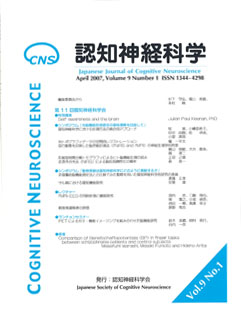 Our review article entitled "Simultaneous fMRI/EEG recording system for sleep study" has been published in Cognitive Neuroscience. Vol.9 No.1, 2007.
 Our review article entitled "Simultaneous fMRI and sleep EEG recording system" has been published in Clinical Electroencephalography. Vol.47 No.6, 2007.