情報通信研究機構 未来ICT研究所
情報通信研究機構 未来ICT研究所
覚醒時に比べ傾眠時では自発性脳活動ネットワークの結合特性が変化し情報処理の効率性が低下することを "Efficiency of a "Small-World" Brain Network Depends on Consciousness Level: A Resting-State fMRI Study" というタイトルで、Cerebral Cortexに発表した。
article link
概要:It has been revealed that spontaneous coherent brain activity during rest, measured by functional magnetic resonance imaging (fMRI), self-organizes a “small-world” network by which the human brain could sustain higher communication efficiency across global brain regions with lower energy consumption. However, the state-dependent dynamics of the network, especially the dependency on the conscious state, remain poorly understood. In this study, we conducted simultaneous electroencephalographic recording with resting-state fMRI to explore whether functional network organization reflects differences in the conscious state between an awake state and stage 1 sleep. We then evaluated whole-brain functional network properties with fine spatial resolution (3781 regions of interest) using graph theoretical analysis. We found that the efficiency of the functional network evaluated by path length decreased not only at the global level, but also in several specific regions depending on the conscious state. Furthermore, almost two-thirds of nodes that showed a significant decrease in nodal efficiency during stage 1 sleep were categorized as the default-mode network. These results suggest that brain functional network organizations are dynamically optimized for a higher level of information integration in the fully conscious awake state, and that the default-mode network plays a pivotal role in information integration for maintaining conscious awareness.
この論文の内容が神戸新聞にて紹介されました。
神戸新聞2013年2月23日朝刊
『眠いとなぜミス増える? 脳機能低下の仕組みを解明』
※ Firefoxをお使いの場合、表示されるPDF文書が非常に見にくい場合があります。その場合は、オプションメニューからPDFの表示方法を "Adobe Acrobatを利用(Firefoxe内で表示)" に変更してください。
浅いノンレム睡眠、深いノンレム睡眠、レム睡眠におけるdefault-mode networkの違いを "Connectivity pattern changes in default-mode network with deep non-REM and REM sleep" というタイトルで、Neuroscience Reseachに発表した。
article link
_Cover.png) 概要:Recent studies have compared default-mode network (DMN) connectivity in different arousal levels to investigate the relationship between consciousness and DMN. The comparison between the DMN in rapid eye movement (REM) sleep with that in non-REM (NREM) sleep is useful for revealing the relationship between arousal level and DMN, because the arousal level is at its lowest during deep NREM, while during REM sleep it is as high as wakefulness. Functional magnetic resonance imaging (fMRI) and polysomnogram data were acquired from participants in REM, deep NREM, and light NREM sleep, and the DMN was compared using functional connectivity analysis. Our analysis revealed that functional connectivity among the DMN core regions -- the posterior cingulate cortex, rostral anterior cingulate cortex, and inferior parietal lobule -- remained consistent across sleep states. In contrast, connectivity involving the DMN subsystems of REM sleep differs from that of NREM sleep, and the change well accounts for the characteristics of REM sleep. Our results suggest that both the DMN core region and subsystems may not relate to the maintenance of arousal. The DMN core network and subsystems may respectively serve to integrate brain regions and perform function specific to each level of arousal.
概要:Recent studies have compared default-mode network (DMN) connectivity in different arousal levels to investigate the relationship between consciousness and DMN. The comparison between the DMN in rapid eye movement (REM) sleep with that in non-REM (NREM) sleep is useful for revealing the relationship between arousal level and DMN, because the arousal level is at its lowest during deep NREM, while during REM sleep it is as high as wakefulness. Functional magnetic resonance imaging (fMRI) and polysomnogram data were acquired from participants in REM, deep NREM, and light NREM sleep, and the DMN was compared using functional connectivity analysis. Our analysis revealed that functional connectivity among the DMN core regions -- the posterior cingulate cortex, rostral anterior cingulate cortex, and inferior parietal lobule -- remained consistent across sleep states. In contrast, connectivity involving the DMN subsystems of REM sleep differs from that of NREM sleep, and the change well accounts for the characteristics of REM sleep. Our results suggest that both the DMN core region and subsystems may not relate to the maintenance of arousal. The DMN core network and subsystems may respectively serve to integrate brain regions and perform function specific to each level of arousal.
K-complexに伴うfMRI信号変化と心拍変動との関係を 「自発性K複合に伴うfMRI信号変化と心拍変動との関係 −EEG/fMRI同時計測を用いた検討−」 というタイトルで、臨床神経生理学誌に発表した。
概要:健常者18名に対して睡眠ポリグラフとfMRIの同時計測をおこない,自発性K複合の出現に伴うfMRI信号の変化を調べた。また,K複合に付随する一過性の心拍変動とfMRI信号変化との関連性を検討した。その結果,K複合の出現に伴う有意なfMRI信号の増加を小脳,腹側視床,脳幹背側部,側脳室周囲白質および脳室(大大脳静脈槽)で認めた。一方,有意なfMRI信号の減少は右半球島皮質前部で見られた。さらに,K複合に伴うfMRI信号の増加の程度は,付随する心拍変動の大きさに比例していた。以上の結果は,K複合の出現に伴う一過性の心拍変動が特定の脳部位におけるfMRI信号変化に影響を及ぼしうることを示す。また,K複合と自律神経活動の密接な関連や循環調節への自律神経系の関与を考えれば,今回の結果は,睡眠中における自律神経系を介した全身および脳内の循環調節への自発性K複合の関与を示唆するものと思われる。
レム睡眠中の急速眼球運動に伴う脳活動を "Human brain activity time-locked to rapid eye movements during REM sleep" というタイトルで、Experimental Brain Research に発表した。
article link (open access)
概要:To identify the neural substrate of rapid eye movements (REMs) during REM sleep in humans, we conducted simultaneous functional magnetic resonance imaging (fMRI) and polysomnographic recording during REM sleep. Event-related fMRI analysis time-locked to the occurrence of REMs revealed that the pontine tegmentum, ventroposterior thalamus, primary visual cortex, putamen and limbic areas (the anterior cingulate, parahippocampal gyrus and amygdala) were activated in association with REMs. A control experiment during which subjects made self-paced saccades in total darkness showed no activation in the visual cortex. The REM-related activation of the primary visual cortex without visual input from the retina provides neural evidence for the existence of human ponto-geniculo-occipital waves (PGO waves) and a link between REMs and dreaming. Furthermore, the time-course analysis of blood oxygenation level-dependent responses indicated that the activation of the pontine tegmentum, ventroposterior thalamus and primary visual cortex started before the occurrence of REMs. On the other hand, the activation of the putamen and limbic areas accompanied REMs. The activation of the parahippocampal gyrus and amygdala simultaneously with REMs suggests that REMs and/or their generating mechanism are not merely an epiphenomenon of PGO waves, but may be linked to the triggering activation of these areas.
MRマグネット内で閉眼中の被験者の眼位を赤外線カメラの画像から推定する方法を、"Eye Position Estimation During Sleep Using Infrared Video in Functional MRI" というタイトルで、Journal of Advanced Computational Intelligence and Intelligent Informatics, 12(1): 32-40 (2008) に発表した。
(兵庫県立大学・小橋研究室との共同研究)
article link
概要:The determination of eye position during sleep attracted much attention in sleep study. Simulaneous measurement consisting of functional magnetic resonance imaging and infrared video was developed to study the relationship between eye movement and brain function during sleep. Conventional measurement of the eye position using infrared video images is not applicable to experiments during sleep because of the lack of tracking targets such as the pupil. This paper proposes a novel method for determining the eye position during sleep through infrared video images. This method determines the eye position by comparing intensity profile extracted from an image to intensity profiles generated by the artificial neural network (ANN). Experiments showed that the proposed method detected the eye position of the left eye with an error of 3.56±3.61 (RMSE±SD) pixels and that of the right eye with an error of 4.69±4.73 pixels.
ニューラルネットワークを用いたfMRIデータの新しい解析法を "Application of artificial neural network to fMRI regression analysis." というタイトルで、Neuroimage, 29(2):396-408 (2008) に発表した。
article link
概要:We used an artificial neural network (ANN) to detect correlations between event sequences and fMRI (functional magnetic resonance imaging) signals. The layered feed-forward neural network, given a series of events as inputs and the fMRI signal as a supervised signal, performed a non-linear regression analysis. This type of ANN is capable of approximating any continuous function, and thus this analysis method can detect any fMRI signals that correlated with corresponding events. Because of the flexible nature of ANNs, fitting to autocorrelation noise is a problem in fMRI analyses. We avoided this problem by using cross-validation and an early stopping procedure. The results showed that the ANN could detect various responses with different time courses. The simulation analysis also indicated an additional advantage of ANN over non-parametric methods in detecting parametrically modulated responses, i.e., it can detect various types of parametric modulations without a priori assumptions. The ANN regression analysis is therefore beneficial for exploratory fMRI analyses in detecting continuous changes in responses modulated by changes in input values.
第16回認知神経科学会学術集会にて、「fMRI−脳波の同時計測とresting-state fMRI −fMRI による『脳の状態』の計測−」を発表した。
概要:これまでのfMRIは、刺激やタスクに伴って生じる一過性の脳活動の局在を高い空間分解能で同定するための非侵襲的計測法として用いられてきた。そこでは対照条件としてのRest条件では、(1) 明確な脳活動が存在せず、(2) ほぼ一定の状態が保たれている、という暗黙の前提があった。どちらの前提も必ずしも満たされているとは限らない事は以前から知られてはいたが、あくまで実験計画及び解析の際の留意事項にすぎなかった。言い方を変えれば、fMRIでは自発性脳活動はノイズと見なされて研究対象としては扱われてこなかった。しかし、fMRIの実験中に被験者が眠気を感じない事の方が稀であるし、時には実験中に眠ってしまう事も少なくない(睡眠は健常者における最大の脳活動変化である)。また安静時及び睡眠時の脳活動が単に脳のアイドリングではない事は、Steriadeによる神経生理学的知見や90年代後半より極めて活発に研究が進められた睡眠時のmemory consolidationに関する研究からも明らかである。
近年fMRIを用いた研究でも、白発性脳活動を対象とした研究が飛躍的に増加している。これらの研究は大きく二種類に分けることができる。第一はfMRIと脳波を同時に計測し、脳波の基礎律動の変化(=覚醒水準の変動)及び自発性脳活動に伴うBOLD信号の変化から、脳波の基礎律動及び自発性脳活動に関与する脳領域を同定し、その生理学的意義を検討する研究である。第二は、安静覚醒時のfMRI時系列データを用いて、領域間のfMRI信号の相関から機能的結合性 (functionalconnectivity) を計算して、全脳の自発性活動を複数の領域が協調して活動する数種類のネットワークに分類し、健常者と神経・精神疾患患者での相違や意識との関連を検討する研究(ressting-state fMRI) である。そしてこれらの自発性脳活動ネットワークの中核的存在として考えられているのが DefaultMode Network (DMN) である。
本講演では、われわれが行ってきた研究(レム睡眠時の急速眼球運動に伴う脳活動及び覚醒~睡眠に伴うDMNの変動)を紹介しながら、上記の二種類の研究について概観し、今後の仏1RIによる脳研究の方向性を検討-する。
第13回日本ヒト脳機能マッピング学会にて、「EEG/fMRI同時計測を用いたレム睡眠中の急速眼球運動に伴う脳幹部の活動の検討」を発表した。
【目的】EEG/fMRI同時計測を用いた先行研究によって、レム睡眠中の急速眼球運動 (Rapid Eye Movements: REMs) に伴う大脳および間脳の活動が報告されている。一方、REMsに密接に関連するponto-geniculo-occipital (PGO) waveの発生部位である脳幹部の活動については一致した結論は得られていない。その原因として、先行研究で用いられている1.5T MRIのSN比の低さが考えられる。そこで本研究では、レム睡眠中の急速眼球運動に伴う脳幹部の活動を、3T MRIを用いて検討した。 【方法】健常成人8名を対象として、夜間、被験者の睡眠中にfMRIと睡眠ポリグラムの同時計測をおこなった。レム睡眠期およびREMsの開始時点を視察で判定した後、REMsをイベントとしたevent-related analysisをおこなって、レム睡眠中のREMsに伴う脳活動部位を検討した。 【結果】8名の被験者のうち6名がレム睡眠まで到達し、そのうち4名の被験者でREMsに伴うfMRI信号の有意な増加を一次視覚皮質および橋で認めた。一方、REMsに伴うfMRI信号の有意な低下は見られなかった。REMsに伴う橋の活動は、4名の被験者のうち3名の被験者において、橋網様体の位置する橋の正中背側部で一致して見られた。 【考察】本研究により、一貫したREMsに伴う橋網様体の活動がはじめて明らかになった。橋網様体は、REMsの出現と密接に関連するPGO waveの発生部位として知られている。その一方で、橋網様体の活動が覚醒時の視運動性眼振やニコチン誘発性眼振によって生じるとの報告もある。したがって、レム睡眠中のREMsに伴う橋の活動は、REMsの発生と密接に関連するPGO waveを反映するもの、あるいは覚醒時と同様に衝動性眼球運動の実行に関与するものと思われる。
第26回日本生体磁気学会のシンポジウム『機能的MRIによる安静時脳機能ネットワーク: その基盤と展望』において、「Resting-state fMRIによる自発的脳活動の検討と睡眠時脳活動への応用」を発表した。
Resting-state fMRIによる自発的脳活動の検討と睡眠時脳活動への応用
概要: 近年、課題負荷や刺激提示をおこなわず安静状態での自発的脳活動を計測するresting-state fMRIと脳部位間のfMRI信号の相関を基にした機能的結合解析を用いて、脳の自発的活動によって形成される特定の脳部位間でのネットワークの存在が明らかになるとともに、脳の自発的活動ネットワークと認知能力や意識、またアルツハイマー病などの精神神経疾患との関連に注目が集まっている。Resting-state fMRIと機能的結合解析による脳内ネットワークの検討は、その神経基盤や解釈などで検討すべき課題も多いが、睡眠中や麻酔下あるいは昏睡状態など課題遂行が困難な状態での脳活動および脳機能を調べる上で有用である可能性がある。 最近、我々は健常成人を対象に睡眠時の脳活動をEEGとfMRIを用いて同時に測定し、睡眠段階によるdefault modenetworkを中心とした脳内ネットワークの差異について検討をおこなった。その結果、default mode network内での変化は見られないものの、default modenetworkとその他の部位との機能的結合が睡眠段階によって変化することが明らかになった。本発表では、resting-state fMRIを用いた自発性脳内ネットワークの検討法について、その概略を紹介するとともに、上記の研究について詳しく報告し、resting-statefMRIを用いて自発性脳活動を検討する上で留意すべき点などについて議論したい。
Neuroscience 2010 (San Diego, CA, USA にて、"Activation of the pontine reticular formation accompanying rapid eye movements during REM sleep: A high-resolution fMRI study" を発表した。
Activation of the pontine reticular formation accompanying rapid eye movements during REM sleep: A high-resolution fMRI study.
概要: Numerous animal studies have demonstrated that the brain stem plays a key role in the generation of ponto-geniculo-occipital (PGO) waves and rapid eye movements (REMs) during REM sleep, but the related neural substrate in the generation of these phenomena so far remains unclear in humans. In recent years, we and other groups have reported that several brain areas, such as the thalamus and the visual cortex, are activated accompanying REMs by using fMRI. Among these studies, activation areas in the visual cortex and the thalamus mostly overlap; however activation regions in the brain stem are inconsistent. We investigated brain stem activities accompanying REMs by a high-resolution fMRI at 3T to precisely localize activation regions. Five healthy adults participated in the study. We recorded fMRI and EEG data simultaneously while they slept in an MR scanner. After subtracting scanning and pulse artifacts from the EEG, we visually identified the periods of REM sleep and onsets of REMs from the EEG, EOG and video recordings of participants’ eyelid movements. Then we conducted event-related analysis with SPM8. As reported in previous studies, we observed REMs-related activation of the visual cortex and the thalamus. Note that we found additional activation specially localized in the medial part of the pontine tegmentum; likely the pontine reticular formation (PRF). It is well known that the PRF, as well as the pedunculopontine nucleus, is closely related in the generation of PGO waves; however, we assume that the activation of the PRF is associated with the generation of REMs, based on the following findings: (1) activation of the PRF was observed during horizontal optokinetic nystagmus (OKN) in a previous study; (2) a lesion in this area impairs REMs as well as waking saccades. Although our result, consequently, suggests that REMs and waking saccades share a neural substrate underlying oculomotor function, this does not mean REMs are completely equivalent to waking saccades. In our additional control experiment we were not able to observe pronounced pontine activation elicited by horizontal OKN or waking saccades, even when we adopted block design, which has higher detection power than event-related design. In addition, a previous study on cats reported that the slope of main sequence amplitude-velocity relationship of REMs is higher than that of waking saccades, implying that the oculomotor system is in a different state of excitation during REM sleep and wakefulness. Considering these findings, our result suggests that the PRF has a different functional significance in REMs during REM sleep and saccades during wakefulness.
第29回 国際臨床神経生理学会のシンポジウム "Rapid eye movements (REM) and sleep: EEG and fMRI study" にて、"fMRI activation time-locked to rapid eye movements during REM sleep" を発表した。
fMRI activation time-locked to rapid eye movements during REM sleep.
概要: Frequent eye movement and high incidence of vivid dreams are characteristic of rapid eye movement (REM) sleep. Several studies have reported REM-related neural activities in cortical and subcortical areas, which are inactive during waking saccades in total darkness. However, the exact locations of these areas are still unclear. To identify the neural substrate of rapid eye movements during REM sleep in humans, we conducted simultaneous functional magnetic resonance imaging (fMRI) and polysomnographic recording during REM sleep. Event-related fMRI analysis time-locked to the occurrence of REMs revealed that the activation of the pontine tegmentum, ventroposterior thalamus, primary visual cortex (V1), putamen, and limbic areas (the anterior cingulate, parahippocampal gyrus, and amygdala) occur in association with REMs. The results of a control experiment during which subjects made self-paced saccades in total darkness showed no activation in the visual cortex. The time-course analysis of blood oxygenation leveldependent (BOLD) responses indicated that the activation of the pontine tegmentum, ventroposterior thalamus, and primary visual cortex started before the occurrence of REMs (from 3 to 5 s). In contrast, the activations of the putamen and limbic areas accompanied REMs. The results can be summarized as follows: (1) the REM-related activation of the pontine tegmentum, thalamus, and primary visual cortex without visual input from the retina provides neural evidence for the existence of ponto-geniculo-occipital waves (PGO-waves) in humans; (2) the V1 activation prior to REM is consistent with the scanning hypothesis of dreaming; i.e., dream imagery occurs first, and then the eyes scan it; and (3) the activation of the parahippocampal gyrus and amygdala accompanying REMs implies that REMs essentially function as triggers that activate these memory-related areas rather than mere epiphenomenon of PGO-waves.
第29回 国際臨床神経生理学会にて、"The reticular activating system is associated with spontaneous fluctuations of alpha rhythm: a simultaneous EEG/fMRI study" を発表した。
The reticular activating system is associated with spontaneous fluctuations of alpha rhythm: a simultaneous EEG/fMRI study.
概要:Objective: In recent years, brain activation correlated with spontaneous fluctuations of alpha rhythm during relaxed wakefulness has been intensively investigated using the simultaneous EEG/fMRI recording technique. Whereas a negative correlation between alpha rhythm and BOLD signal in the occipital cortices was consistently observed, positive and negative correlations in other regions are controversial among previous studies. Furthermore, studies that investigated activation of subcortical regions related to alpha rhythm are scarce. In this study, we investigated activation of subcortical structures, related to spontaneous alpha rhythm during resting state, besides cortical regions. Methods: We simultaneously measured EEG and fMRI data from 26 participants (20-35 year-old). During 10-minutes recording sessions, they lay supine in a 3T-MRI scanner and were resting with eye closed. After removing the MR gradient artifacts and the ballistocardiogram artifacts in the EEG data, we calculated relative alpha power (ratio between 8-12 Hz and 1-12 Hz band power) corresponding to each EPI image from 7.5 sec windows of EEG data, which was used as a regressor of interest in individual analysis. In addition, we used realign parameter as a nuisance regressor. Individual and group analysis was done using SPM2. Results: We found a negative correlation in the occipital cortex, which is consistent with previous studies. We also found a negative correlation in the superior temporal gyrus and the sensorimotor cortex. In contrast to the negative correlation, a positive correlation was mainly observed in subcortical regions, such as the thalamus, lateral part of the midbrain tegmentum and medial part of the pontine tegmentum. Conclusions: We found specific subcortical structures related to spontaneous fluctuations of alpha rhythm. These structures seem to correspond to the reticular activating system. Our finding, thus, suggests that reticular activating system is involved in fluctuations of spontaneous alpha rhythm during resting wakefulness.
第33回 日本神経科学大会にて、「アルファ波に関連する脳部位の同定におけるcanonical HRFの畳み込みの影響」を発表した。
Influence of convolution of the hemodynamic response function on identification of alpha-fMRI correlations
概要:Objective: Recently, brain activation correlated with spontaneous fluctuations of alpha rhythm during wakefulness has been intensively investigated. Almost these studies used canonical hemodynamic response function (HRF); however, this is not justified yet. To investigate influence of convoluting HRF, we compared results of the following two models in this study: the first model used alpha time series, and the second model used that convolved with HRF.Methods: We simultaneously measured EEG and fMRI data from 26 participants (age: 20-35 year-old). During 10-minutes recording sessions, they lay supine in a 3T-MRI scanner and were resting with eye closed. We calculated relative alpha power (ratio between 8-12 Hz and 1-12 Hz band power) corresponding to each EPI image from 7.5-sec window of EEG data, which was used as a regressor of interest. For the second model, we used another regressor derived from the alpha time series convolved with HRF. In addition, we used realign parameter as a nuisance regressor in both models. Individual and group analysis was done using SPM2.Results: For the first model, we found positive correlation in the thalamus, the midbrain-pontine tegmentum and the cerebellum. We also found negative correlation in the occipital cortex, the sensorimotor cortex and the superior temporal gyrus. In contrast to the first model, we did not observe positive correlation, and we observed negative correlation almost all cortical regions, except for prefrontal areas, for the second model.Conclusions: We demonstrated that convolution of HRF largely affects detection of alpha-fMRI correlations. In contrast to the first model, results of the second model, especially positive correlation, is artifactual, because positive correlation was observed only in the lateral ventricle. Our results clearly show that we must consider validity of convoluting HRF in the future studies exploring neural correlates of spontaneous fluctuations of alpha rhythm.
Neuroscience 2009 (Chicago, IL, USA) にて "Heart rate fluctuation affects fMRI signals across the cerebral cortices during REM and light non-REM sleep, but not during deep non-REM sleep" および "Alterations in brain network corresponding to the fluctuations in arousal level within a short time period" を発表した。
Heart rate fluctuation affects fMRI signals across the cerebral cortices during REM and light non-REM sleep, but not during deep non-REM sleep
概要:Recently, the effects of cardiac activity, especially heart rate, on fMRI signals have been reported. We previously reported a positive correlation between heart rate and fMRI signals across many cortical areas, including the medial prefrontal and occipital cortices, during light non-REM sleep (sleep stage 2). During light non-REM sleep, a biphasic change in the heart rate occurs accompanied by a K-complex which is a characteristic waveform in sleep EEG. Thus, we assumed that the positive correlation between the 2 parameters may be light non-REM sleep-specific. To test this assumption, we investigated the relationship between the 2 parameters by performing correlation (simple regression) analysis using SPM2 during different sleep stages: REM and light and deep non-REM sleep (sleep stage 3/4). We simultaneously obtained fMRI and polysomnography recordings in 15 participants (age, 22-31 years) while they slept during 2 consecutive nights. Of the 15 participants, 12 entered REM sleep, and 11 and 12 showed stable light and deep non-REM sleep, respectively. In REM sleep, as well as light non-REM sleep, significant positive correlation between the 2 parameters was observed across many cortical areas, including the posterior cingulate and posterior insular cortices. The positive correlation was also observed in both the posterior thalamus and cerebellum. In contrast, in deep non-REM sleep, significant positive correlation was observed only in the occipital cortex and thalamus. Moreover, the positive correlation in the thalamus observed during deep non-REM sleep was significantly higher than that observed during light non-REM and REM sleep. Negative correlation was not observed during any of the sleep stages. Heart rate fluctuation is relatively greater in light non-REM and REM sleep than that in deep non-REM sleep. In addition, the coefficient of variation of R-R intervals (CVRR) at each sleep stage also showed the same tendency in our study. Thus, the positive correlation between the 2 parameters across the cerebral cortices may be due to the greater heart rate fluctuation. On the other hand, the positive correlation in the thalamus may be related to the observed correlation between EEG delta power and regional cerebral blood flow in the thalamus, because the thalamus is associated with the regulation of cardiovascular activity and the generation of EEG delta rhythm. In conclusion, the relationship between heart rate and fMRI signals differs across each sleep stage. Hence, the effect of heart rate fluctuation during sleep should be considered while conducting an fMRI study..
Alterations in brain network corresponding to the fluctuations in arousal level within a short time period
概要:[PURPOSE] Several studies have examined the characteristics and spatial patterns of the default mode network (DMN) during resting wakefulness. Arousal levels do not remain constant even for short periods of time. Even when participants claim to have a clear sensorium while being scanned, the electorencephalogram (EEG) pattern indicates that high- and low-arousal microstates alternate with each other. In this study, we attempted to elucidate whether the activity of the DMN brain network fluctuates with changes in arousal level during a relatively short time period.
[METHOD] We acquired simultaneous functional magnetic resonance imaging (fMRI) and EEG data from 12 study participants who were required to stay awake and keep their eyes closed while being scanned within the MRI scanner. The arousal level_defined as the ratio between EEG power, rAT = (EEG power in alpha-band (8-12 Hz) / the EEG power in the theta-band (4-8 Hz)) was observed to have significantly fluctuated during the session (10 min). Higher rAT values indicate higher arousal levels. We defined microstates with a rAT>1 as “high-arousal microstates,” and microstates with a rAT<=1 as “low-arousal microstates.” Subsequently, using preprocessed fMRI data, we evaluated functional connectivity during high- and low-arousal microstates. Functional connectivity was calculated across 22 regions of interest (ROIs) including subcortical regions, visual cortices, and fundamental elements of DMN using Pearson’s correlation coefficient. Thereafter, we tested whether the strength of connectivity varied between high- and low-arousal microstates by using Wilcoxon’s matched-pair signed-rank test. [RESULTS and DISCUSSION] First, the positive connectivity between the fundamental elements of the DMN was observed; it was observed that the strength of connectivity significantly increased during the high-arousal microstate as compared to the low-arousal (p < 0.001). To our knowledge, this is the first study showing that DMN characteristics change with short-time fluctuations in the arousal level. In addition, we found that the strength of the anticorrelation between the thalamus and the visual cortex increased during the high-arousal compared to the low-arousal microstates (p = 0.002); positive connectivity within the visual cortex was enhanced during the high-arousal microstates (p = 0.02). These modulations associated with the thalamo-visual network appears to be consistent with the evidence that the EEG alpha-wave originates from the thalamo-visuo circuit. The present study indicates that the functioning of brain networks varies with fluctuations in the arousal levels in resting wakefulness.
第32回 日本神経科学大会にて、「REM睡眠中の眼球運動は覚醒時の眼球運動に関わるネットワークを異なる仕方で利用している」および「皮質でみられる睡眠時に特異的な心拍変動とfMRI信号の相関」を発表した
REM睡眠中の眼球運動は覚醒時の眼球運動に関わるネットワークを異なる仕方で利用している
概要:Using functional magnetic resonance imaging (fMRI), we explored functionally connected brain networks associated with waking saccades (visually guided saccade, VG; self-paced saccade with eyes open/closed; EO/EC) and rapid eye movements (REMs) during REM sleep.Functional connectivity between V1 and saccade-related areas were significantly positive in VG and EO, and almost no connectivity was observed in EC cases. The positive connectivity between V1 and saccade-related areas represents the mechanism for controlling eye movements on the basis of visual information.In the case of REMs, robust but negative connectivity was observed between V1 and saccade-related areas. The results suggest that the same network for the control of eye movements is associated with both waking saccades with visual information processing and REMs; however, the functional role of the network during REM sleep may be different from that during performing waking saccade tasks.
皮質でみられる睡眠時に特異的な心拍変動とfMRI信号の相関
概要:We previously reported a positive correlation between heart rate and fMRI signal across many cortical areas including the medial prefrontal and the occipital cortex during light non-REM sleep. To examine whether this correlation is sleep-specific, we performed investigations at relatively high- and low-arousal wakefulness. In high-arousal wakefulness, in contrast to light non-REM sleep, only negative correlation was observed in the periventricular area. The negative correlation was preserved in low-arousal wakefulness. This result clearly shows that the positive correlation across the cortex is sleep-specific. In light non-REM sleep, K-complex occurs spontaneously or provocatively accompanied by a biphasic change in the heart rate. Thus, such a change in heart rate accompanied by the K-complex may lead, at least partially, to the sleep-specific positive correlation between heart rate and the fMRI signal across the cortex.
第11回 日本ヒト脳機能マッピング学会にて、「短時間での覚醒レベルの揺らぎに対応した脳内ネットワークの変動」および「側脳室周囲でみられるfMRI信号に及ぼす心拍の影響」を発表した。
短時間での覚醒レベルの揺らぎに対応した脳内ネットワークの変動
概要:【目的】覚醒レベルの指標として脳波のAlpha帯域とTheta帯域のパワーの比(A/T比)が用いられている。高いA/T比は覚醒レベルが高いことをあらわす。覚醒状態であってもA/T比の値には数秒単位での揺らぎが存在すること、すなわち覚醒レベルには短期的な揺らぎがあることが知られている。本研究では複数脳領域間でのBOLD信号間の機能的結合の変化を算出することにより、短時間での覚醒レベルの揺らぎに対応して脳内ネットワークの変動が起こっているかを調べた。【方法】対象は健常成人12名。安静閉眼状態で、10分間のEEG-fMRI同時記録をおこなった。SPM5およびMATLABでfMRIデータを前処理し、複数の関心領域 (ROI) の時系列信号を算出した。同時記録されたEEGからA/T比を算出し、郭10分のセッション内をA/T比が1以上の区間と1未満の区間とにする。それに基づき、高A/T比と低A/T比の区間でのROI間の機能的結合の変化を比較した。A/T比の変動が著しく少ないデータは除外し、計39セッションのデータを用いた【結果】複数の機能的結合は、A/T比の高低に対応して有意な変動を示した。A/T比(覚醒レベル)が高い区間ではそれが低い区間と比較して、default mode network (DMN) と呼ばれるネットワーク内 (p<0.001)、および一次視覚野 (V1) 内での結びつきの強さが強くなる (p=0.018) ことが明らかになった。また、視床-V1間での負の機能的結合は、A/T比が高い場合に強まった (p=0.018)。【結論】これまでの先行研究では、DMNは覚醒レベルの変動に拠らずに安定して存在すると考えられてきた。しかし本研究は、A/T比で定義された短時間での覚醒レベルの揺らぎに対応してDMNの活動も揺らいでいることを初めて明らかにした。また高いA/T比に対応して見られる視床と一次系を含むネットワークの変化は、Alpha波の生成メカニズムを反映している可能性がある。これらのネットワークが覚醒の揺らぎに果たしている役割は、今後の検討課題である。
側脳室周囲でみられるfMRI信号に及ぼす心拍の影響
概要:【目的】近年、被験者の心拍変動に伴って機能的MRI(fMRI)信号が変化することが報告されている。しかし、心拍がfMRI信号に及ぼす影響が、どのような要因によって生じるかは明らかではない。本研究では、fMRIと心電図の同時計測をおこない、心拍とfMRI信号との相関関係がみられる脳内部位を同定して、心拍がfMRI信号に影響を及ぼすメカニズムを解明しようと試みた。
【方法】対象は健常成人14名。安静閉眼状態で、10分間のfMRI撮像をおこなった。fMRIデータの解析にはSPM2を用い、解析結果を各被験者のT1およびT2*強調画像に重畳して、心拍とfMRI信号の有意な相関がみられた部位と解剖学的構造との対応を調べた。
【結果】心拍とfMRI信号との有意な正の相関が脳幹部でみられた。一方、心拍とfMRI信号との有意な負の相関は側脳室周囲でみられた。個々の被験者で、相関関係を示した部位と解剖学的構造との対応を検討したところ、有意な相関がみられたすべての被験者で共通して、負の相関がみられた部位は上衣下静脈など側脳室周囲を走行する静脈と一致していた。だが、正の相関を示す部位は、脳幹部を除き、特定の構造との対応はみられなかった。
【考察】脳幹部でみられた心拍とfMRI信号の正の相関は、先行研究で報告されているとおり、脳底部を走行する動脈内の心拍変動に伴う血流量変化に起因すると考えられる。一方、負の相関を示す側脳室周囲の静脈には深部白質の髄質静脈が流入すること、深部白質は動脈支配の境界に位置すること、また栄養する血管は灰白質とは異なることから、心拍とfMRI信号との負の相関は深部白質に特異的な循環調節メカニズムが関与するものと考えられる。さらに、今回併せて報告するnon-REM睡眠中にみられた一過性の心拍変動に伴う側脳室周囲でのfMRI信号変化も、部位の一致を踏まえれば、同様のメカニズムが関与しているものと考えられる。
Neuroscience 2008 (Washington DC, USA) にて "Exploring changes in the brain network related to human sleep states using graph theory" を発表した。
概要:The focus of our research is on changes in the functional organization of the human brain during the course of REM/NREM sleep. In particular, we examined what brain regions are indispensable for sleep-related network, and how the brain network switches in association with REM/NREM transition. Given several lines of evidence showing the possibility that REM/NREM sleep have different role in information processing, e.g. memory consolidation, there would be at least two different brain networks corresponding to REM/NREM sleep. In order to evaluate switching of brain network, we employed graph theory, which is usually considered as the most appropriate framework to analyze complex networks such as human brain network. We recorded fMRI on thirteen normal subjects while they slept in an MR scanner without any goal-directed tasks. Sleep stages were scored from EEG, EMG, EOG, and video data which were simultaneously recorded with fMRI. First, we modeled the sleeping brain as the sleeping brain as a non-directed weighted graph with continuous arch weights given by the hyperbolic correlation measure between spontaneous BOLD signals at any two different regions of interests. Next, we calculated the vulnerability of the network to identify what brain regions are the most indispensable to the functionally connected brain networks. During REM sleep states, following brain regions were detected as vulnerable regions: the thalamus, putamen, amygdala, and insula cortex. During NREM sleep (SWS; slow-wave-sleep), the most central areas are: the thalamus, putamen, amygdale, and caudal part of hippocampus. Finally, in order to evaluate which brain regions communicate with the vulnerable regions, we evaluated the network efficiency. As a result, it is revealed that hippocampus interacted with association area (BA46, 19, 9, and precuneus) during REM sleep, while hippocampus dominantly communicates with subcortical areas (the thalamus, putamen, amygdale, insula, and caudate) during NREM sleep. The thalamus significantly exchanged information between primary cortices (V1, S1, A1, and M1) only during REM sleep. Other vulnerable regions, i.e. the putamen, amygdala, and insula, equally interacted with cortical areas during both REM and NREM. The result of our experiment is likely the first evidence to clearly show the switching of the functionally connected brain network accompanying REM/NREM transition. The observed switching of information flow between thalamus/hippocampus and other brain regions is consistent with ‘two stage hypothesis of memory consolidation’ proposed by Stickgold et al. (2000).
第38回 日本臨床神経生理学会・学術大会の日本臨床神経生理学会・日本睡眠学会合同シンポジウム「睡眠研究の進歩」において、「EEG/fMRI同時計測による睡眠研究:K-complexの脳循環自動調節への関与」を発表した。
概要:1930年代にその存在が報告されて以来、数多くの研究によってK-complexの機能や形態的諸特性が明らかにされてきた。その一方で、K-complexの発生や機能に関わる神経基盤については、EEGやMEGを用いた試みでは必ずしも一致した結果は得られていない。また、PETやfMRIなどの脳機能画像法を用いた研究は、これまでほとんどおこなわれていない。そこで我々は、EEG/fMRI同時計測を用いて、K-complexに伴う脳活動部位の同定を試みた。15名の実験協力者の睡眠中にポリソムノグラムとfMRIの同時計測を実施した。視察によってKcomplexを同定した後、事象関連fMRI解析によってK-complexに伴う信号変化が生じる脳部位を調べた。その結果、K-complexに伴うBOLD信号の有意な上昇が側脳室周囲白質と小脳で見られ、視覚野と聴覚野では有意な信号の低下が見られた。しかし、これら部位の信号変化は、同時期の心拍変動とも有意に相関していた。また、K-complex後の心拍変動の大きさと、K-complexに伴う信号変化の程度の間には、正の相関が見られた。K-complexと自律神経活動との関連、特に心拍数や血圧がK-complexの直後に上昇することが、先行研究によって報告されている。また、BOLD信号は脳血流変化に依存したものであり、安静閉眼時では心拍変動がBOLD信号変化に影響を及ぼすとの報告がある。したがって、K-complexに伴うBOLD信号変化は、K-complexを生じさせる神経活動による脳血流変化を反映したものというよりも、K-complex直後の心拍変動による脳内循環の変化を反映したものと考えられる。さらに、K-complexおよび心拍数変化に伴う信号変化を示した側脳室周囲白質の領域は、watershed領域と一致する。Watershed領域は、循環圧低下による虚血を生じやすく、慢性の高血圧などにより脳循環の自動調節が障害されやすい部位として知られている。PETを用いた先行研究では、watershed領域の白質でのみ睡眠中にも自動調節機能が維持されると報告されており、今回示した側脳室周囲白質でのK-complexあるいは心拍変動に伴うBOLD信号の変化には、脳循環の自動調節が関与していると思われる。今回の結果は、K-complexの神経基盤やその機能を示唆するものではない。しかし、K-complexが睡眠中の脳内循環自動調節脳の調整に関与していることを示すものと考えられる。ただし、側脳室周囲白質以外の部位における信号変化も脳循環自動調節で説明できるかどうかは、更なる検討が必要である。また、K-complexの神経基盤の解明には、BOLD信号に対するK-complexに伴う自律神経活動による影響を分離する工夫が求められる。
日本神経回路学会 第18回 全国大会にて、階層型クラスタリングを用いてヒト睡眠状態に特異的な神経回路を示した「階層型クラスタリングを用いたs-BOLD解析によるREM/NREM睡眠ネットワークの解明」を発表した。
概要:Using spontaneous BOLD fluctuation analysis based on hierarchical clustering technique, we tried to identify neural networks corresponding to REM/NREM sleep. We found 2 sub-networks during REM and 3 networks during NREM. The results may suggest different functional roles of REM/NREM.
Human Brain Mapping2008 (Melbourne, Australia) にて spontaneous BOLD analysisにより睡眠段階の移行に伴う視床皮質回路の切り替わりを示した "Dynamic switching of thalamocortical network with transition of human states between NREM and REM sleep" を発表した。
概要:Increased attention has been directed at investigating spontaneous BOLD activities, and these studies have made clear that several brain regions may form state-dependent network without any goal directed tasks, and the spontaneous network is thought to correlate with some brain functions. Several recent studies suggest that human sleep is not only a resting state but a state processing important function, such as memory consolidation, and suggest the possibility that the REM and NREM sleep have different function. Therefore, it is reasonable to suppose that state-dependent network switches its characteristics accompanying sleep stage transition between REM and NREM. In this study, using spontaneous BOLD activity analysis, we tried to find out dynamic switching of state-dependent brain network with transition of sleep state.
REM睡眠とNREM睡眠において、睡眠段階(脳の状態)に応じて神経回路網が切り替わる様子をfMRI-EEG同時計測により明らかにした。その結果を、"Dynamic switching of thalamocortical network in association with transition between REM and NREM sleep" というタイトルで国立生理学研究所において開催された「神経科学の道具としてのfMRI研究会」で口頭発表した。
概要:In recent years, increased attention has been directed at investigating spontaneous brain activities; examination of spontaneous BOLD activity has made clear that several brain regions form a functionally- connected "resting-state network" during wakefulness without goal-directed tasks. Another attention has been paid to brain activities during sleep. Although there seems to be also no explicit goal-directed tasks during human REM/NREM sleep state, several recent studies suggest that spontaneous brain activities during sleep are associated with important brain functions such as memory consolidation. Therefore, considering functionally-connected brain network associated with human REM/NREM sleep may be helpful to investigate sleep-stage-related brain functions. To investigate characteristics of spontaneous networks linked to human sleep states, we used fMRI with simultaneous polysomnographic recording, and examined functional connectivity between thalamus and other cortical areas, using a technique with Spearman's rank-correlation coefficient between the BOLD signals. We found that a set of regions, including DLPFC, anterior cingulate and hippocampus, showed significantly increased connectivity to thalamus during NREM sleep period (sleep stage 3/4). In contrast, during REM sleep which is usually linked to vivid dreaming, we found a thalamocortical network including visual cortices, hippocampus and primary motor area. In addition, only during REM sleep, we found a significant functional connectivity between the thalamus and the posterior cingulate cortex, precuneus, and inferior temporal gyrus; these regions are known as a part of "default network". Our findings show that thalamocortical network switches in association with transition of sleep states between NREM and REM, and suggest that identifying functionally-connected brain network using correlation coefficient between BOLD signals is a useful tool for exploring brain functions of NREM and REM sleep.
日本神経科学大会のテクニカルセミナーにて "Relationship between the EEG rhythm and BOLD signals" を発表した。
概要:Recording fMRI and an EEG simultaneously is effective for studying spontaneous brain activities. We used this method to examine the relationship between an EEG rhythm and a BOLD signal. Some studies have hypothesized that the hemodynamic response for a change in power of certain EEG frequency bands, such as alpha waves, is canonical in shape. However, the appropriate response shape for a change in the rhythmic EEG has not yet been determined. We measured the EEG and fMRI simultaneously while subjects were in a resting or sleeping state. We applied nonlinear regression analysis using an artificial neural network to detect correlations between the changes in rhythmic EEG waves and fMRI signals without a priori hypothesis of the response shape.
REM睡眠中の急速眼球運動に伴う賦活を脳波とfMRIの同時計測によって検討し、12th Annual meeting Human Brain Mapping (Florence, Italy) において発表した。
概要:Despite numerous studies on REM sleep, two issues remain unsettled in this field. One is the lack of clear evidence for PGO-waves in humans; the other is whether there is a relationship between rapid eye movement and dreaming, the so called "scanning hypothesis" according to which REM is directly correlated to the movements of the gaze in a dream scene. Though some EEG/MEG studies found neural activities in the occipital area and the thalamus which accompanied REM, the precise location of these brain areas is still unclear. To address these issues, ten subjects slept in an MR scanner for a few hours in the early morning. We simultaneously obtained an fMRI signal accompanied by polysomnography and video recording of eyelid movements. After removing artifacts caused by the MR scanner from the polysomnograph, we visually identified the period of REM sleep and eye movement onsets using both the polysomnograph and the video recording. Six of the subjects showed stable REM sleep which lasted for 20-40 min. Event-related fMRI analysis was done using SPM2. The expected BOLD response for each REM onset was modeled by a canonical HRF. In addition, other models in which the event onset time was shifted were applied to detect REM-related activities with different latencies. All six of these subjects showed a similar fMRI activation pattern accompanying REM: the pontine tegmentum, thalamus, primary visual cortex, extrastriate cortex, cuneus, cingulated cortex, basal ganglias and cerebellum were significantly activated. The activation of the first three areas probably reflects neural activities caused by PGO-waves. In addition, whereas the activation of the first three areas tended to precede REM onset, the activation of the other areas was delayed by a few seconds. The precedent activation to REM in V1 suggests that dream imagery occurs first, and then eyes move to scan the image as is consistent with the scanning hypothesis.
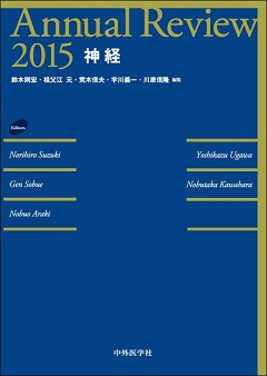 Annual Review 2015 神経に、fMRIと脳波の同時計測法による睡眠時脳活動および意識に関する研究の解説『脳波とfMRIの同時計測による睡眠と意識の研究』を寄稿
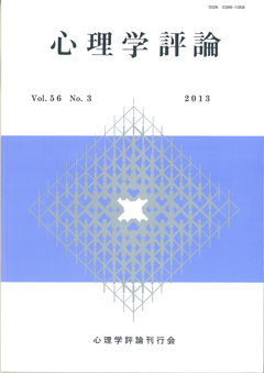 心理学評論 Vol.56 No.3に、非侵襲脳機能計測法の解説『脳を測る ―改訂 ヒトの脳機能の非侵襲的測定―』を寄稿
※ 著者原稿が こちら からご覧いただけます。
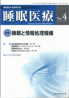 睡眠医療 Vol.4 No.4 特集「睡眠と情報処理機構」に、fMRIと脳波の同時計測法による睡眠時脳活動の研究の解説『fMRIを用いた睡眠中の自発性および誘発性脳活動の検討』を寄稿
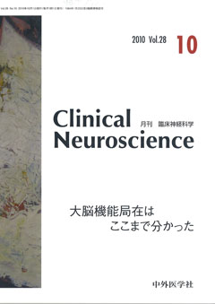 Clinical Neuroscience 2010年10月号 特集「大脳の機能局在はここまで分かった」に、機能的磁気共鳴画像(fMRI)を用いた機能的結合解析についての解説 『自発性脳活動が示す脳部位間の機能的結合』を寄稿
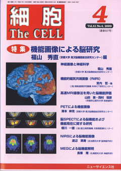 細胞 2009年4月号 特集「機能画像を用いた脳研究」に、 機能的磁気共鳴画像(fMRI)を用いた脳研究の解説 『機能的磁気共鳴画像(fMRI)』 を寄稿
 睡眠学(日本睡眠学会 編)に、機能的磁気共鳴画像(fMRI)を用いた睡眠研究の解説 『fMRIによる睡眠研究』 を寄稿
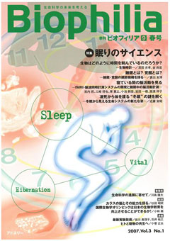 Biophilia 2007年9月号 特集「眠りのサイエンス」に、fMRIと脳波の同時計測を用いた睡眠時脳活動についての研究の解説 『寝ている間の脳活動を見る:fMRI-脳波同時計測システムの開発と睡眠中の脳活動計測』 を寄稿
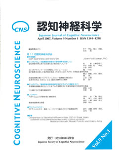 認知神経科学 2007年4月号に、fMRIと脳波の同時計測を用いた睡眠研究についての解説 『fMRI-EEGの同時計測と睡眠研究』 を寄稿
 臨床脳波 2005年6月号 特集「律動脳波」に、当チームが開発したEEG、fMRIおよび眼球運動の同時観察・計測システムを用いた睡眠研究についての解説 『睡眠時脳波とfMRIの同時計測』 を寄稿