Strategic Basic Research Programs CREST
[Biodynamics] Creation of Fundamental Technologies for Understanding and Control of Biosystem Dynamics
Research agenda
"Cell-cell adhesion・Elucidation of the mechanism of order formation of the skeleton and development of epithelial barrier manipulation technology"
Research period:October 2013 - March 2018
Principal Investigator:Tsukiko TSUKITA(Professor,Graduate School of Frontier Biosciences, Osaka University)
Research contributor : Kazuhiro OIWA
Outline /
Purpose /
Methods /
Results
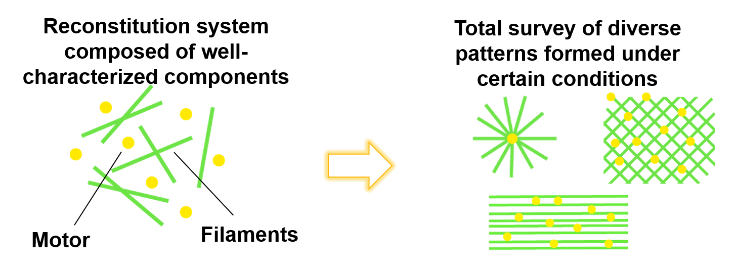
Exhaustive search for structures that are created by mixing microtubules and protein motors

Methods
After a detailed analysis of the minor processes resulting from the mixing of microtubules and proteins motor or microtubule-associated proteins, the process of forming a global order structure that is actively formed by mixing them at different ratios is described.

Contents of Research and Results
①Construction of a wide area 3 d fluorescent molecule imaging system
Considering the dynamic ordering of microtubule networks ranging from 2 to 3 dimensional, I. e., a wide range of structural formation, we introduced a confocal optical microscope system as a fast observable fluorescence microscope with a wide viewing, high-resolution and pseudo- 3 d fluorescence system.
This was the first successful observation of a detailed observation of the formation process of the mesososcopic dynamic vortex structure of the cm size, which was created by the sliding motion of microtubules and dyneins.
。
②Construction of in vitro reconstruction system by a known element combination for verification of mathematical model
Predicted by the theoretical model of Ishihara G.
The opening angle consisting of 2 microtubules extending from the active complex is one of the important parameters of the ordering.
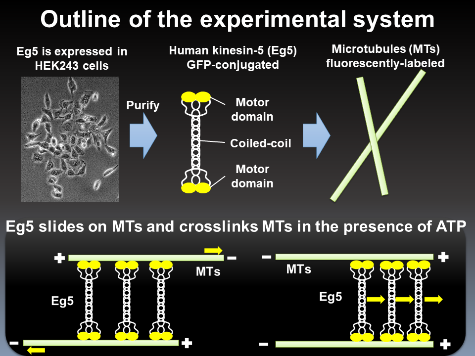
To provide precise control of this, we attempted to reconstruct the microtubule network as a polymerized nucleus with a fragment of Chlamydomonas flagellar axoneme and tubulin.
It was confirmed that the element model with the polarity of microtubules was constructed, and the microtubule network was dynamically changed by the addition of kinesin beads, but the difficulty of parameter setting due to the heterogeneity of the axon fragment was clarified, and the examination object was shifted to the reconstitution system using only known materials.
Results→


